 IJCP Editorial Team
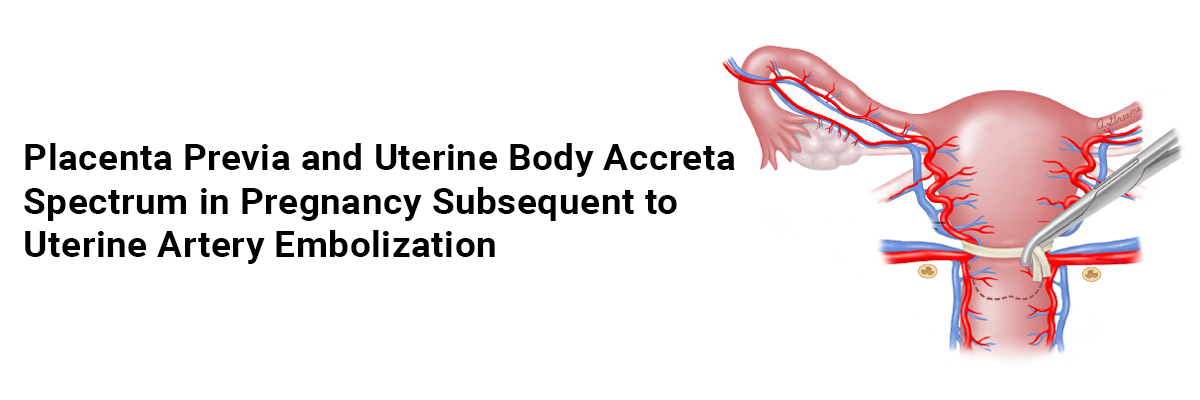
Placenta previa and uterine body accreta spectrum in pregnancy subsequent to uterine artery embolization
A report describes a case of a 35-year-old woman, gravida 2 para 1, who had undergone bilateral Uterine artery embolization (UAE) with a gelatin sponge for postpartum hemorrhage (PPH) due to uterine atony following vaginal delivery in her previous pregnancy. She documented no UAE-related complications, with a resumption of menstruation, and subsequently conceived naturally. However, she learned to have a placenta previa.
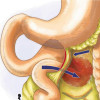
An ultrasonography and MRI examination at 33 weeks gestation showed a placenta on the anterior to the lateral wall of the uterus, consistent with total placenta previa. The patient displayed no evidence of placenta accreta spectrum (PAS) in the lower uterine segment.
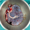
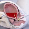
The patient underwent a cesarean section at 37 weeks of gestation due to placenta previa and delivered a healthy female infant (with birth weight 2906 g, Apgar score 8 at 1 min) by an oblique incision in the lower uterine body to bypass the placenta in the maximum possible way. The placenta self-detached from the lower uterine segment but not from the uterine body. Thus, the surgeons manually separated the placenta and diagnosed the patient clinically with uterine body placenta accreta spectrum (PAS) with placenta previa. Her uterus did not contract, and the bleeding persisted. Thus, she received an intrauterine balloon tamponade device with 100 mL of sterile liquid and a suture of the uterus. Her bleeding finally stopped, and the surgery was finished with a total blood loss of 2940 mL.
After one h 30 min, the patient experienced 1040 mL of bleeding from the drainage tube of the intrauterine balloon tamponade device. At this time, her systolic blood pressure was 119 mmHg, pulse rate was 76 beats/min, with stable hemodynamics. However, her uterus did not show any contractions, and active bleeding persisted. She did not desire to preserve her fertility and had developed an allergic reaction to the contrast media during her previous UAE. Thus, she underwent a subtotal hysterectomy instead of UAE. The surgery was uneventful, with a total blood loss of 145 mL. The patient received eight units of red blood cells and six units of fresh-frozen plasma during the procedure.
Her Pathological report suggested a diagnosis of placenta accreta since the chorionic villi were in direct contact with the myometrium. The patient displayed good postoperative condition and received discharge seven days after surgery.
Sugai S, Yamawaki K, Haino K., et al. Unexpected uterine body placenta accreta spectrum with placenta previa in a subsequent pregnancy after uterine artery embolization: a case report. BMC Pregnancy Childbirth. 2022; 22. https://doi.org/10.1186/s12884-022-05031-0
IJCP Editorial Team
Comprising seasoned professionals and experts from the medical field, the IJCP editorial team is dedicated to delivering timely and accurate content and thriving to provide attention-grabbing information for the readers. What sets them apart are their diverse expertise, spanning academia, research, and clinical practice, and their dedication to upholding the highest standards of quality and integrity. With a wealth of experience and a commitment to excellence, the IJCP editorial team strives to provide valuable perspectives, the latest trends, and in-depth analyses across various medical domains, all in a way that keeps you interested and engaged.
More FAQs by IJCP Editorial Team


Recent FAQs

Related FAQs


Medtalks is India's fastest growing Healthcare Learning and Patient Education Platform designed and developed to help doctors and other medical professionals to cater educational and training needs and to discover, discuss and learn the latest and best practices across 100+ medical specialties. Also find India Healthcare Latest Health News & Updates on the India Healthcare at Medtalks
Please login to comment on this article