 IJCP Editorial Team
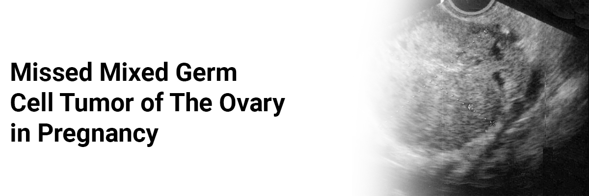
Missed mixed germ cell tumor of the ovary in pregnancy
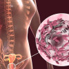
A report describes a case of a 34-year-old primigravida at 37 weeks + 5 days POG who came in latent labor. A term scan demonstrated a fibroid of 11x 9 cm in the posterior wall in the lower uterine segment with the central cystic area.
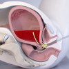
The patient underwent emergency LSCS and delivered a male baby (wt 2.005 kg). Exteriorization of the uterus revealed a 10 x 8 cm necrotic mass posterior to the uterus in rectovaginal space, which was inferred as FIGO Stage IC1 ovarian carcinoma.
Histopathology of the mass demonstrated mixed germ cell tumor: 60% yolk sac tumor and 40% dysgerminoma.
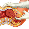
MRI done after two weeks showed a lesion of 10.4 x 9.5 x 7.3 cm in the rectovaginal pouch, compressing and displacing the rectum posteriorly and to the left side, also displacing the uterus and bladder anteriorly. Her Left ovary was normal, while her Right ovary was not visualized. Therefore, she received four cycles of chemo comprising cisplatin and etoposide.
After chemo, a PET scan revealed a lesion of 6.7x 6.2 x 4.2 cm in the rectovaginal area just right to the midline, with the right ovary not seen distinctly. Hence, she underwent laparoscopic right oophorectomy. Histopathology showed profound regressive changes with few scattered atypical cells with no definitive evidence of residual tumor. Follow-up serum LDH and AFP came within normal limits, and the patient was advised to follow up once in 3 months to look for any recurrences.
Pais J A, Fernandes V R, Rao S V, A case of missed mixed germ cell tumor of ovary in pregnancy. Indian J ObstetGynecol Res. 2023;10(2):214-216
IJCP Editorial Team
Comprising seasoned professionals and experts from the medical field, the IJCP editorial team is dedicated to delivering timely and accurate content and thriving to provide attention-grabbing information for the readers. What sets them apart are their diverse expertise, spanning academia, research, and clinical practice, and their dedication to upholding the highest standards of quality and integrity. With a wealth of experience and a commitment to excellence, the IJCP editorial team strives to provide valuable perspectives, the latest trends, and in-depth analyses across various medical domains, all in a way that keeps you interested and engaged.
More FAQs by IJCP Editorial Team


Recent FAQs

Related FAQs


Medtalks is India's fastest growing Healthcare Learning and Patient Education Platform designed and developed to help doctors and other medical professionals to cater educational and training needs and to discover, discuss and learn the latest and best practices across 100+ medical specialties. Also find India Healthcare Latest Health News & Updates on the India Healthcare at Medtalks
Please login to comment on this article