 IJCP Editorial Team
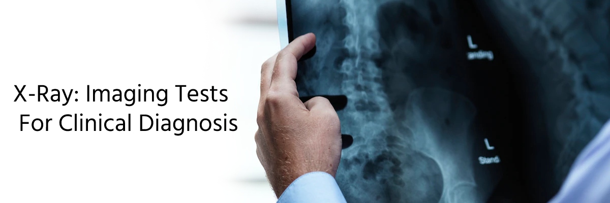
X-Ray: Imaging Tests For Clinical Diagnosis
X-rays are a type of electromagnetic wave radiation used in medical imaging procedures to examine the human body's internal structures. It is a quick, painless test wherein X-ray beams passed through the body give an image output that is studied for clinical investigations. In this procedure, an x-ray beam is passed through the body by an x-ray machine. The x-ray striking the internal structures is either absorbed or scattered in different amounts. The undeflected x-rays are transmitted to a detector and processed to generate an image on a film or computer screen. The rate of absorbency of the radiation varies for different body parts depending on the density. Dense materials like bones appear white as calcium absorbs the maximum x-rays. Soft tissues and fat absorb less and thus appear gray. Air absorbs the least, generating a black image on X-ray film or digital media.
Types of X-ray Studies
Though the basic principle is similar, medical imaging procedures are classified into several types, each employing a unique set of technologies and techniques, serving varied purposes. These include:
● Radiography: a single image for later analysis.
● Mammography: used for imaging the internal structures of the breasts.
● Fluoroscopy: allows for the real-time monitoring of surgery or the movement of a contrast agent (also known as a "dye") through the body by displaying continuous x-ray images on a monitor. It can result in comparatively significant radiation doses, particularly for complex interventional procedures (such as inserting stents or other devices inside the body) that call for prolonged fluoroscopy exposure.
● Computed Tomography: as the images are reconstructed from many individual x-ray projections, CT requires a higher radiation dose than conventional radiography.
Application of X-ray procedure
Standard X-rays have a wide range of applications, such as detecting chest conditions like pneumonia, diagnosing tumors, and locating bone diseases or injuries. X-ray technology is also used in other diagnostic procedures, such as computed tomography (CT) scans, arteriograms, and fluoroscopy. An X-ray may be recommended for the following conditions:
If the doctor suspects a fracture.
To determine the cause of pain and swelling in certain body parts.
To detect structural problems in bones, joints, or soft tissues.
To carry out routine screenings for cancer and other diseases.
Advantages and Disadvantages of X-Rays
X-ray imaging tests are widely accepted as useful medical tools for various examinations and procedures. The wide acceptance depends on varied factors like:
It is non-invasive.
It is a painless procedure.
It aids in medical and surgical treatment planning.
It guides medical professionals as they place stents, catheters, or other devices to treat tumors, remove blood clots, or clear other blockages.
X-rays can be carried out safely on all ages, including infants.
However, there are certain risks associated with using X-rays:
Excessive exposure to x-rays can lead to the development of cancer later in life
It may cause cataracts and hair loss
There is an increased possibility of adverse reactions to an intravenously injected contrast agent.
If performed during pregnancy, it may have detrimental effects on the fetus.
In the balance of benefits and risks of using x-rays, it is crucial to consider the following factors:
Younger patients are more susceptible to radiation; special precautions should be taken to limit radiation exposure to the pediatric population during all X-ray imaging exams.
Due to the potential effects of radiation exposure on the developing fetus, special precautions should be taken when imaging pregnant patients.
The benefit of detecting disease should be carefully weighed against the risks of imaging screening studies on healthy, asymptomatic patients.
Some organs are more radiosensitive than others in the body.
The risk of developing cancer from exposure to medical imaging radiation is generally very low, and it is dependent on the following:
1. Radiation dose - The higher the dose, the greater the lifetime risk of cancer.
2. Age of the patient - A patient who receives X-rays at a younger age has a higher lifetime risk of cancer than one who receives them at an older age.
3. Sex - Women are slightly at more risk of developing radiation-associated cancer than their male counterparts
Tips to prepare for an X-ray
Upraise your healthcare provider about your medical history, allergies and medications, if any.
Inform the doctor if you're pregnant or breastfeeding.
Wear comfortable or loose-fitting cloths
You may be asked to change into a gown before the X-ray' if necessary.
For X-rays other than bone, you should:
Avoid using products such as creams, lotions, or perfume.
Remove metal objects like jewelry and hairpins as they may interfere with the imaging process
Do not eat or drink several hours beforehand (for GI X-rays).
IJCP Editorial Team
Comprising seasoned professionals and experts from the medical field, the IJCP editorial team is dedicated to delivering timely and accurate content and thriving to provide attention-grabbing information for the readers. What sets them apart are their diverse expertise, spanning academia, research, and clinical practice, and their dedication to upholding the highest standards of quality and integrity. With a wealth of experience and a commitment to excellence, the IJCP editorial team strives to provide valuable perspectives, the latest trends, and in-depth analyses across various medical domains, all in a way that keeps you interested and engaged.
More FAQs by IJCP Editorial Team


.png)


Medtalks is India's fastest growing Healthcare Learning and Patient Education Platform designed and developed to help doctors and other medical professionals to cater educational and training needs and to discover, discuss and learn the latest and best practices across 100+ medical specialties. Also find India Healthcare Latest Health News & Updates on the India Healthcare at Medtalks
Please login to comment on this article