 IJCP Editorial Team
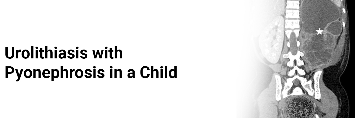
Urolithiasis with pyonephrosis in a child
A report describes a case of a 10-year-old boy who initially presented to the emergency department with right abdominal pain, throbbing in nature, associated with nausea, vomiting, and loose stool for two days.
Examination revealed a palpable mass over the right hypochondriac region for which he received conservative treatment for acute appendicitis. However, an ultrasound showed gross hydronephrosis secondary to pelvic-ureteric junction obstruction. CT abdomen showed right nephrolithiasis and vesicoureteric calculus with right hydroureter and gross hydronephrosis, which could be secondary to the right vesicoureteric junction or pelvic-ureteric junction stricture. This prompted an urgent surgical intervention with retrograde pyelography (RPG) and ureteroscopy (URS) by inserting a ureteral stent under general anesthesia. The patient subsequently received a discharge to home.
However, three days after discharge, the patient returned with a complaint of fever, hematuria, and dysuria. He received treatment for right pyelonephritis. However, after five days, his condition did not improve despite the high dose of antibiotics. Examination revealed him febrile with a temperature of 39.9, Blood pressure of 110/70 mmHg, pulse rate of 134 beats per minute, and pain score of 4/10.
Abdominal examination demonstrated a ballotable right kidney and positive right renal punch. Blood analyses showed worsening white cell count and a high C-reactive protein (CRP). Urine full examination and microscopic examination (UFEME) demonstrated nitrates, leucocytes, hematuria, and proteinuria. Urine culture and sensitivity (C&S) revealed an extended-spectrum beta-lactamase (ESBL) organism, pseudomonas aeruginosa. Abdominal X-ray showed a ureter in-situ stent and two clusters of multiple small radio-opaque lesions over the right renal, representing nephrolithiasis.
Ultrasound abdomen revealed right hydronephrosis, slightly less than the previous study, and internal echo within the dilated system that may represent pyonephrosis. Based on the urine culture result, he received antibiotic meropenem 20 mg/kg which improved his clinical and blood parameters after 48 hours. After completing meropenem for seven days, the patient received a discharge with oral ciprofloxacin 250 mg twice daily for three weeks for better treatment coverage because of the presence of ESBL organism in the urine previously. In this case, the final diagnosis was right pyonephrosis with extended-spectrum beta-lactamase pseudomonas aeruginosa with underlying right nephrolithiasis and vesicoureteric calculus.
Md Sabudin SN, Yaacob LH, Draman N. An atypical presentation of urolithiasis with pyonephrosis in a child: A case report. Electron J Gen Med. 2023;20(2):em457. https://doi.org/10.29333/ejgm/12852
IJCP Editorial Team
Comprising seasoned professionals and experts from the medical field, the IJCP editorial team is dedicated to delivering timely and accurate content and thriving to provide attention-grabbing information for the readers. What sets them apart are their diverse expertise, spanning academia, research, and clinical practice, and their dedication to upholding the highest standards of quality and integrity. With a wealth of experience and a commitment to excellence, the IJCP editorial team strives to provide valuable perspectives, the latest trends, and in-depth analyses across various medical domains, all in a way that keeps you interested and engaged.
More FAQs by IJCP Editorial Team


Recent FAQs


Related FAQs


Medtalks is India's fastest growing Healthcare Learning and Patient Education Platform designed and developed to help doctors and other medical professionals to cater educational and training needs and to discover, discuss and learn the latest and best practices across 100+ medical specialties. Also find India Healthcare Latest Health News & Updates on the India Healthcare at Medtalks
Please login to comment on this article