 IJCP Editorial Team
Successful Vaginal Delivery following External Cephalic Version in a Woman with a Large Partial Uterine Septum
A recent report describes a case of an 18-year-old nulligravida who initially presented with a complaint of 2 years of infertility and a known uterine septum. She revealed significant gynecological history for menarche at the age of 13, regular monthly menses, and a history of treated chlamydia. She also documented a history of congenital pulmonary stenosis treated with two angioplasties and hypertension managed with losartan.
Her most recent echocardiogram revealed an ejection fraction of 60-65% and no significant valvular disease.
A pelvic exam revealed an anteverted, 8-week size uterus. Transvaginal ultrasound showed a uterus measuring depth with normal appearing ovaries.
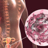
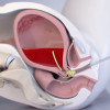
3D transvaginal ultrasound revealed the uterine septum with a 2 cm deep indentation with an angle of 65°. A hysterosalpingogram revealed two uterine cornua with normal appearing and patent fallopian tubes. Her Laboratory studies were normal.
She reported her first pregnancy spontaneously two months after the hysterosalpingogram. Part of the uterine septum was visible on an 18-week obstetrical ultrasound performed in this first pregnancy. During her first pregnancy, she received the diagnosis of preeclampsia with severe features at 37 weeks of gestation.
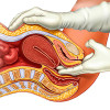
An ECV, followed by labor induction, resulted in an uncomplicated vaginal delivery.
The patient then documented her second pregnancy spontaneously four months after her first delivery. She then presented as a 20-year-old gravida 2 para 1001 at 37 weeks and six days of gestational age with confirmed breech presentation and a posterior placenta. The estimated fetal weight by ultrasound came as 3003 grams, and the maximum vertical amniotic fluid pocket was 5.7 cm.
At 38 weeks and 0 days, the patient underwent ECV, which was successful on the second attempt, and the patient received anti-D immune globulin after the procedure.
After the ECV, the patient received a discharge and was scheduled to return for induction of labor at 39 weeks 0 days. On returning for labor induction, she again displayed a breech position by ultrasound. So she again underwent an epidural and ECV. During labor, the patient was diagnosed with preeclampsia with severe blood pressure and started on magnesium sulfate for seizure prophylaxis.
She delivered a singleton male in cephalic position vaginally at 39 weeks two days of gestation, weighing 3510 grams with Apgar scores of 8 at 1 minute and nine at 5 minutes.
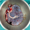
After the fetus and placenta were delivered, the patient displayed retained membranes that did not deliver with gentle traction. Manual uterine exploration disclosed membranes firmly adhered in the right cornu, which was presumed to have contracted around the membranes holding them in place. The membranes were expelled manually with traction. Manual uterine exploration afterward showed a thick uterine septum confirming the previous 3D TVUS and hysterosalpingogram findings.
The patient displayed an uncomplicated postpartum course and received another dose of anti-D immune globin on postpartum day two since the infant was Rh-positive. The mother and infant both received a discharge on postpartum day 2.
Park KE, et al. Successful Vaginal Delivery after External Cephalic Version in a Woman with a Large Partial Uterine Septum. Case Reports in Obstetrics and Gynecology. 2021; 2021. https://doi.org/10.1155/2021/9912271
IJCP Editorial Team
Comprising seasoned professionals and experts from the medical field, the IJCP editorial team is dedicated to delivering timely and accurate content and thriving to provide attention-grabbing information for the readers. What sets them apart are their diverse expertise, spanning academia, research, and clinical practice, and their dedication to upholding the highest standards of quality and integrity. With a wealth of experience and a commitment to excellence, the IJCP editorial team strives to provide valuable perspectives, the latest trends, and in-depth analyses across various medical domains, all in a way that keeps you interested and engaged.
More FAQs by IJCP Editorial Team


Recent FAQs

Related FAQs


Medtalks is India's fastest growing Healthcare Learning and Patient Education Platform designed and developed to help doctors and other medical professionals to cater educational and training needs and to discover, discuss and learn the latest and best practices across 100+ medical specialties. Also find India Healthcare Latest Health News & Updates on the India Healthcare at Medtalks
Please login to comment on this article