
 IJCP Editorial Team
Management of breech presentation with a large pelvic hydatid cyst in late pregnancy
A report describes a case of a 19-year-old woman, gravida 2, para 1, admitted at 37 weeks of gestation with breech presentation. She suffered pregnancy complications due to multiple hepatic hydatids (the largest cyst: 6.0*4.3 cm) and a large pelvic hydatid cyst (10.0*8.2 cm).
Fetal ultrasound revealed the term of the fetus as approximately 37 weeks [biparietal diameter (BPD): 8.9 cm, abdominal circumference (AC): 31.7 cm, femur length (FL): 67 cm]. Three weeks before admission, the patient felt distended and presented at the hospital, which was her first antenatal examination during this pregnancy. An obstetric ultrasound revealed hepatic and pelvic hydatids. The size of the pelvic hydatid cyst remained the same over these weeks (9.5*8.6 cm). She reported delivering her first baby at home three years earlier. She also reported being screened for local endemic diseases and found to have hepatic and abdominopelvic hydatid disease and underwent surgical treatment two years ago. However, a lack of conscientiousness for medical care and her remote location refrained her from getting regular reexaminations or medication.
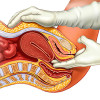
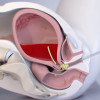
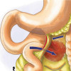
Ultrasound showed the location of the pelvic hydatid cyst to be posterior to the uterus as a bulge in the posterior fornix, which was confirmed by Vaginal and rectal examinations. Her other laboratory results came out to be normal.
The woman declined a Caesarean section; thus, a management plan according to her situation was formulated. Since a large cyst might direct labor obstruction and abnormal fetal presentation and be more prone to rupture in late pregnancy, the surgeons drained the cyst through the posterior fornix under the guidance of transabdominal ultrasound (draining out approximately 400 ml of liquid). After the disappearnce of the cyst, they monitored the patient carefully for half of the day, concerning bleeding, fetal movement, and uterine contraction. After confirming the patient was free of complications, the surgeons successfully performed an external cephalic version. They discharged the patient after two days of observation, as she showed no signs of labor.
One week later, the woman delivered a baby boy naturally with a cephalic presentation and received a proposal to undertake medical management for HCD follow-up.
Li P, Wang Y, Yang Q. et al. Management of breech presentation with a large pelvic hydatid cyst in late pregnancy in Tibet: a case report. BMC Pregnancy Childbirth. 2022;22. https://doi.org/10.1186/s12884-022-05180-2
IJCP Editorial Team
Comprising seasoned professionals and experts from the medical field, the IJCP editorial team is dedicated to delivering timely and accurate content and thriving to provide attention-grabbing information for the readers. What sets them apart are their diverse expertise, spanning academia, research, and clinical practice, and their dedication to upholding the highest standards of quality and integrity. With a wealth of experience and a commitment to excellence, the IJCP editorial team strives to provide valuable perspectives, the latest trends, and in-depth analyses across various medical domains, all in a way that keeps you interested and engaged.
More FAQs by IJCP Editorial Team


Recent FAQs
Related FAQs


Medtalks is India's fastest growing Healthcare Learning and Patient Education Platform designed and developed to help doctors and other medical professionals to cater educational and training needs and to discover, discuss and learn the latest and best practices across 100+ medical specialties. Also find India Healthcare Latest Health News & Updates on the India Healthcare at Medtalks
Please login to comment on this article