 IJCP Editorial Team
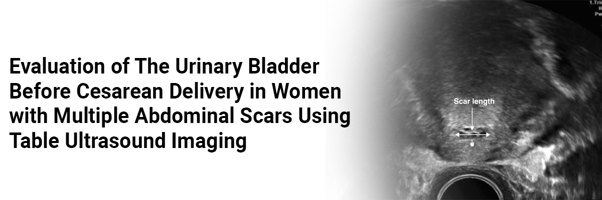
Evaluation of the Urinary Bladder Before Cesarean Delivery in Women with Multiple Abdominal Scars using Table Ultrasound Imaging
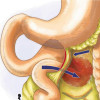
The present report describes a case of a 36-year-old primipara admitted at 39 weeks of gestation for labor induction to indicate a small-for-gestational-age fetus and hypertension in pregnancy. The patient underwent three previous surgical procedures: an open midline laparotomy for perforated appendicitis, a laparoscopic adhesiolysis for an adhesive ileus, and another laparoscopic procedure as part of fertility treatment. She reported having difficulty emptying her bladder; however, she received no previous urogynecology assessment. Given the previous abdominal operations, the surgeons performed abdominal sonography of the mother to map the bladder anatomy, which revealed an abnormally high-situated urinary bladder extending up to the level of the umbilicus. The fetus gained a cephalic position with an estimated weight of 2900 g and three loops of the nuchal cord. After discussion, induction of labor was chosen.
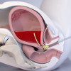
Before labor induction, the surgeons took measures to minimize the risks of bladder injury by identifying the upper limit and outlines of the urinary bladder by transabdominal ultrasound with a longitudinal midline view. During labor, a suspicious CTG prompted performing cesarean delivery (CD).
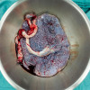
A repeat transabdominal ultrasound scan performed in the theater on the operating table before cleaning and draping showed that even after bladder emptying, the upper limit of the urinary bladder extended up to the level of the umbilicus on ultrasound. The surgeons used a longitudinal midline incision and entered the peritoneum sub umbilically through a small window. The bladder appeared to obstruct access to the lower uterine segment. They mobilized the bladder into the pelvis and small intestine adhesions caudally. They longitudinally incised the anterior uterine segment because the access to the lower uterine segment remained obstructed, increasing the risk for potential bladder injury. Finally, they delivered the baby via reverse breech and repaired the uterine incision in 2 layers with interrupted sutures. Retrograde filling of the bladder with methylene blue excluded the unrecognized bladder injuries. The patient was stable after the procedure.
Fangmann L-C, Henrich W, Hinkson L. Assessment of the urinary bladder prior to cesarean delivery in women with multiple abdominal scars through operation table ultrasonography: a case report. AJOG Global Reports. 2023; 3(1). https://doi.org/10.1016/j.xagr.2022.100138.
IJCP Editorial Team
Comprising seasoned professionals and experts from the medical field, the IJCP editorial team is dedicated to delivering timely and accurate content and thriving to provide attention-grabbing information for the readers. What sets them apart are their diverse expertise, spanning academia, research, and clinical practice, and their dedication to upholding the highest standards of quality and integrity. With a wealth of experience and a commitment to excellence, the IJCP editorial team strives to provide valuable perspectives, the latest trends, and in-depth analyses across various medical domains, all in a way that keeps you interested and engaged.
More FAQs by IJCP Editorial Team


Recent FAQs

Related FAQs


Medtalks is India's fastest growing Healthcare Learning and Patient Education Platform designed and developed to help doctors and other medical professionals to cater educational and training needs and to discover, discuss and learn the latest and best practices across 100+ medical specialties. Also find India Healthcare Latest Health News & Updates on the India Healthcare at Medtalks
Please login to comment on this article