 IJCP Editorial Team
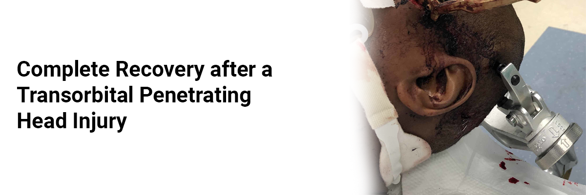
Complete recovery after a transorbital penetrating head injury
|
Computed
tomography (CT) |
intraparenchymal
hemorrhage in the right frontal basal lobe |
|
CT
angiography (CTA) |
Associated
linear tract extending from the posterior aspect of the hematoma to the left
parietal lobe through the genu of the corpus callosum, left globus pallidus,
anterior limb of the internal capsule, and left posterior insula, as well as
a right orbital roof fracture and focal subarachnoid hemorrhage (SAH) |
IJCP Editorial Team
Comprising seasoned professionals and experts from the medical field, the IJCP editorial team is dedicated to delivering timely and accurate content and thriving to provide attention-grabbing information for the readers. What sets them apart are their diverse expertise, spanning academia, research, and clinical practice, and their dedication to upholding the highest standards of quality and integrity. With a wealth of experience and a commitment to excellence, the IJCP editorial team strives to provide valuable perspectives, the latest trends, and in-depth analyses across various medical domains, all in a way that keeps you interested and engaged.
More FAQs by IJCP Editorial Team


Recent FAQs


Related FAQs


Medtalks is India's fastest growing Healthcare Learning and Patient Education Platform designed and developed to help doctors and other medical professionals to cater educational and training needs and to discover, discuss and learn the latest and best practices across 100+ medical specialties. Also find India Healthcare Latest Health News & Updates on the India Healthcare at Medtalks
Please login to comment on this article