Sonographic Morphology of the Pubic Symphysis in Pregnancy: Correlations with Fetal and Maternal Parameters
A recent prospective observational study by Wozniak et al., published in the Journal of Clinical Medicine, investigated the anatomical changes of the pubic symphysis (PS) during different stages of pregnancy using standard ultrasonography.1 The study aimed to define measurable parameters of the PS and explore their correlations with fetal and maternal anthropometric characteristics.
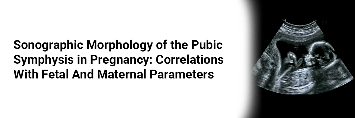
The research enrolled 225 pregnant women aged 23–41 years and measured key sonographic parameters: pubic symphysis width (PSw), depth (PSd), intertubercular distance (PT-PT), and a newly defined metric—pubic symphysis entry middle width (PSemw). These values were correlated with fetal weight, maternal age, pre-pregnancy weight, and weight gain during pregnancy.
The findings revealed that PSw (range: 2.2–11.3 mm) was the most consistent parameter, while PSd (range: 5.4–22.6 mm) showed the greatest variability. PSemw and PSw were most strongly correlated with fetal weight (r = 0.28 and 0.29, respectively; p < 0.001), while PT-PT was significantly associated with maternal age (p = 0.03). Both PSemw and PSw increased significantly with maternal weight and gestational weight gain (p < 0.001). Notably, stratification by fetal weight demonstrated that PSemw and PSw increased significantly in fetuses >3000 g compared to those ≤1000 g or in the 1001–2000 g range (p = 0.02). Additionally, nulliparous women carrying fetuses ≤2000 g exhibited significantly larger PT-PT distances than multiparous counterparts (p = 0.03), highlighting the influence of parity on pelvic morphology.
The study introduces the term "PS entry" to describe a trapezoidal area above the PS disc, bordered by the pubic tubercles and the superior border of the PS disc—an easily visualized and measurable region via ultrasound. Clinically, these findings offer practical reference values for obstetricians. Standard values reported were: PSemw = 6.5 ± 3.4 mm, PSw = 6.4 ± 2.9 mm, PSd = 14.8 ± 4.8 mm, and PT-PT = 15.8 ± 5.3 mm. Deviations from these values may indicate abnormal pelvic adaptation or an increased risk of conditions such as diastasis.
In conclusion, the study underscores fetal weight and maternal weight as primary drivers of sonographic PS changes during pregnancy. These insights can enhance diagnostic accuracy, inform delivery planning, and prompt future longitudinal studies assessing PS changes across trimesters and postpartum recovery. The reproducibility of measurements and the introduction of a novel anatomical reference significantly contribute to obstetric ultrasound practice.
(Source: Wozniak S, Piatek A, Kurc-Darak B, Domagala Z, Paulsen F, Florjanski J. Sonographic evaluations of the pubic symphysis at different stages of pregnancy. J Clin Med. 2025;14(11):3898. doi:10.3390/jcm14113898. Available from: https://www.mdpi.com/2077-0383/14/11/3898 )


Recent FAQs


Related FAQs


Medtalks is India's fastest growing Healthcare Learning and Patient Education Platform designed and developed to help doctors and other medical professionals to cater educational and training needs and to discover, discuss and learn the latest and best practices across 100+ medical specialties. Also find India Healthcare Latest Health News & Updates on the India Healthcare at Medtalks
Please login to comment on this article