
 IJCP Editorial Team
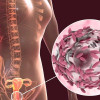
Ovarian adenocarcinoma in a 14-year-old girl
A report describes a case of a 14-year-old girl with ovarian mucinous adenocarcinoma who complained of lower abdominal pain for around 3 weeks and presented with a doubt of an 8 cm left ovarian cyst as revealed by a USG scan.
Her period was regular since menarche at the age of 12 and was earlier diagnosed with albinism.
No abnormality was detected on physical examination, while the ultrasound scan examination of the pelvis showed a cystic mass of 7 x 5.5 cm in the left ovary. MRI images of the abdomen and pelvis too confirmed the USG finding by displaying left ovarian mass with solid and cystic components of 5 x 5 x 7 cm dimension.
Preoperative serum levels according to the ROMA test showed elevation of CA-125: 125.7 IU/mL (normal: < 35 IU/mL) and normal HE4: 36 pmol/L (normal: < 70 pmoI/L). a-fetoprotein (a-FP) was 2.68 ng/mL (normal: 0.79–4.69 ng/mL) and human chorionic gonadotropin (HCG) was < 0.5 mlU/mL. Considering the age of the patients and to avoid invasiveness, laparoscopic surgery was planned.
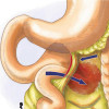
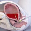
Laparoscopic surgery with the resection of the tumor, left salpingo-oophorectomy and biopsy from the right ovary, was accomplished in oncological purity. Final histology analysis revealed adenocarcinoma of intestinal type of the left ovary without fallopian tube infiltration, but with simple carcinoma cells in peritoneal fluid. No oncological changes were seen in the specimen taken from the right ovary.
Following histopathological reports, the Oncological Council conducted urgent gastroenterological consultation, gastroscopy, colonoscopy and a PET scan, which all did not show any abnormalities.
The results of diagnostic tests prompted the IIB stage of disease according to FIGO 2014. The decision of the European Reference Network for Pediatric Oncology directed six cycles of chemotherapy with an application of carboplatin and paclitaxel. Six months after completing the treatment process, all tumor markers came out to be negative, the control MRI scan was correct and an ultrasonography examination of the pelvis and abdominal was free of any abnormalities. A further follow-up of the patient was recommended.
SOURCE- Drosdzol-Cop A, Mizia-Malarz A, Wilk K, Koszutski T, Wilk K, Stojko F. Ovarian adenocarcinoma in 14-year-old girl. Ginekologia Polska 2022;93(4): 343–344. DOI 10.5603/GP.a2022.0026
IJCP Editorial Team
Comprising seasoned professionals and experts from the medical field, the IJCP editorial team is dedicated to delivering timely and accurate content and thriving to provide attention-grabbing information for the readers. What sets them apart are their diverse expertise, spanning academia, research, and clinical practice, and their dedication to upholding the highest standards of quality and integrity. With a wealth of experience and a commitment to excellence, the IJCP editorial team strives to provide valuable perspectives, the latest trends, and in-depth analyses across various medical domains, all in a way that keeps you interested and engaged.
More FAQs by IJCP Editorial Team


Recent FAQs

Related FAQs


Medtalks is India's fastest growing Healthcare Learning and Patient Education Platform designed and developed to help doctors and other medical professionals to cater educational and training needs and to discover, discuss and learn the latest and best practices across 100+ medical specialties. Also find India Healthcare Latest Health News & Updates on the India Healthcare at Medtalks
Please login to comment on this article