Investigating Placental Abnormalities and Blood Flow in Uteroplacental Insufficiency
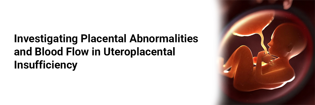
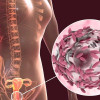
Uteroplacental insufficiency—characterized by inadequate blood flow to the placenta during pregnancy—requires a thorough understanding of the underlying placental pathologies and their clinical significance.
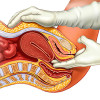
A recent study explored the prevalence, distribution, and types of placental abnormalities in women clinically diagnosed with fetal growth restriction (FGR) and abnormal uterine or umbilical artery Doppler findings. The study analyzed 50 placentae, focusing on both macroscopic and microscopic abnormalities. Key histopathological features examined included infarction, villitis of unknown etiology, intervillous inflammation, and massive perivillous fibrin deposition.
The findings revealed a high frequency of placental pathology among women with uteroplacental insufficiency. Placental infarcts were observed in 98% of cases, villous thrombosis in 94%, intervillous hemorrhage in 98%, and perivillous fibrin deposition in 98%. Inflammatory changes—such as villitis, intervillositis, and deciduitis—were significantly associated with increased resistance in the umbilical artery. Ultrasound assessments showed a 97.8% positive predictive value for detecting placental infarction.
The study concluded that both macroscopic and microscopic placental abnormalities are commonly observed in pregnancies complicated by uteroplacental insufficiency. Notably, conditions such as villitis, intervillositis, deciduitis, and abruption varied significantly between cases with high flow and those with absent or reversed end-diastolic flow (AEDF/REDF). Therefore, histopathological examination of the placenta is recommended postpartum, regardless of the timing of delivery or Doppler findings. However, the study also suggests that inflammatory changes may be more reflective of labor-related events rather than direct indicators of FGR.
Source: Chavan R, Narkhede HR, Satoskar PR, et al. Obst and Gynec India. 2024 Mar 25:1-7.


Recent FAQs


Related FAQs


Medtalks is India's fastest growing Healthcare Learning and Patient Education Platform designed and developed to help doctors and other medical professionals to cater educational and training needs and to discover, discuss and learn the latest and best practices across 100+ medical specialties. Also find India Healthcare Latest Health News & Updates on the India Healthcare at Medtalks
Please login to comment on this article