
 IJCP Editorial Team
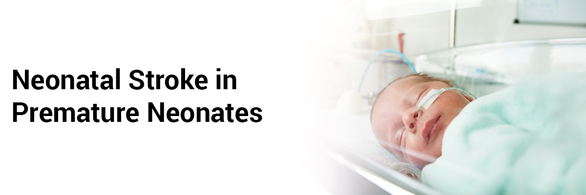
Neonatal stroke in premature neonates
Numerous neuroimaging studies describe the presence of brain lesions in the preterm infant, using cranial ultrasound (cUS) and/or term equivalent age MRI (TEA-MRI). These studies however focus on germinal matrix-intraventricular hemorrhage (GMH-IVH) and white matter injury. Studies regarding perinatal arterial ischemic stroke (PAIS) or cerebral sinovenous thrombosis (CSVT) in the preterm infant are very limited. Several large cohort studies on neuro-imaging in preterm infants do not mention neonatal stroke. The majority of the studies about PAIS exclude preterm infants. Thus a recent study provides an update on neonatal stroke in the preterm infant, with a focus on neuroimaging findings.
Perinatal arterial ischemic stroke-
These centrally located infarcts become apparent after the first postnatal week. They are typically wedge-shaped and mostly do not involve the posterior limb of the internal capsule (PLIC). ‘Cortical sparing’ can be seen in the preterm infant either partial or complete, with complete sparing seen only in infants with a GA<32 weeks and partial sparing in infants with a GA of 33–37 weeks.
Periventricular hemorrhagic infarction
GMH-IVH can convert to PVHI due to impaired venous drainage of the medullary veins into the terminal vein. The area of echogenicity can be globular and continuous with the lateral ventricle and may spread over several lobes, even causing a midline shift. A sagittal plane view can describe the involvement of the trigone. Follow-up scans can reveal the globular lesion evolving into a single cyst communicating with the lateral ventricle (porencephaly) while a triangular-shaped lesion developing multiple cysts which are often not or partly communicating with the lateral ventricle and therefore misdiagnosed as c-PVL.
Neonatal cerebral sinovenous thrombosis Neuroimaging
Neonatal CSVT frequently involves the superficial venous system and can also show thrombosis of multiple sinuses of both the superficial and deep venous system.
Preterm infants often have extensive white matter injury that resembles that of periventricular leukomalacia, which may develop into extensive white matter cysts. This often presents beyond the immediate neonatal period and emerges in combination with late-onset IVH. Thus considering the diagnosis of CSVT in a preterm infant presenting with late-onset white matter injury and unexpected late-onset IVH is important.
Cranial ultrasound
Using Color Doppler cUS can demonstrate partial or a total absence of flow in combination with partial or complete occlusion of the affected sinus(es). Also, cUS can show associated brain lesions in the form of (late-onset) IVH and white matter injury.
Magnetic resonance imaging
Since the symptoms of neonatal CSVT can be non-specific, MR venography (MRV) should also be included in all infants who undergo MRI because of seizures and/or an unusual presentation of (late-onset) IVH or white matter injury.
SOURCE- Steggerda SJ, de Vries LS. Neonatal stroke in premature neonates, Seminars in Perinatology, 2021;45(7). https://doi.org/10.1016/j.semperi.2021.151471.
IJCP Editorial Team
Comprising seasoned professionals and experts from the medical field, the IJCP editorial team is dedicated to delivering timely and accurate content and thriving to provide attention-grabbing information for the readers. What sets them apart are their diverse expertise, spanning academia, research, and clinical practice, and their dedication to upholding the highest standards of quality and integrity. With a wealth of experience and a commitment to excellence, the IJCP editorial team strives to provide valuable perspectives, the latest trends, and in-depth analyses across various medical domains, all in a way that keeps you interested and engaged.
More FAQs by IJCP Editorial Team


Recent FAQs


Related FAQs


Medtalks is India's fastest growing Healthcare Learning and Patient Education Platform designed and developed to help doctors and other medical professionals to cater educational and training needs and to discover, discuss and learn the latest and best practices across 100+ medical specialties. Also find India Healthcare Latest Health News & Updates on the India Healthcare at Medtalks
Please login to comment on this article