 IJCP Editorial Team
Multiple developmental urogenital anomalies - A therapeutic challenge
A report describes a case of a 14-year-old girl who presented with primary amenorrhea and lower abdominal pain with a cyclical increase in severity for three months, without any voiding difficulty.
She consulted a local gynaecologist who recognized hematocolpos and withdrew 100 ml of altered blood by vaginal incision, which temporarily relieved her pain without resuming menstruation. Thus, she approached another physician for further treatment. Clinical examination showed a firm, non-tender, mobile, midline abdominal mass of 10x6 cm of uterine origin. Tanner's stage for secondary sexual characters resembled her age. Local examination (under anaesthesia) demonstrated normal external genitalia, a hypospadiac urethral meatus, absent vaginal lumen without obvious vaginal bulge due to hematocolpos (cryptomenorrhea).
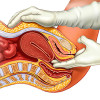
Ultrasound imaging showed a 12cm x 4cm mass indicating hematocolpos and an unascended right kidney. Intravenous Urography confirmed the unascended right kidney in midline at L3 – L4 level. It also showed a duplex collecting system with malrotation on the left side. Both ureters seemed grossly normal, and bladder contour indicated extrinsic compression due to pelvic mass. Micturating cystourethrogram showed a grade IV VUR on the left side. MR urogram identified the following-
- Posteroinferior bladder diverticulum of 37.9mm x 13.8mm with left ureter opening into it.
- Distal left ureteric diverticulum of 14.9mm x 13.6mm.
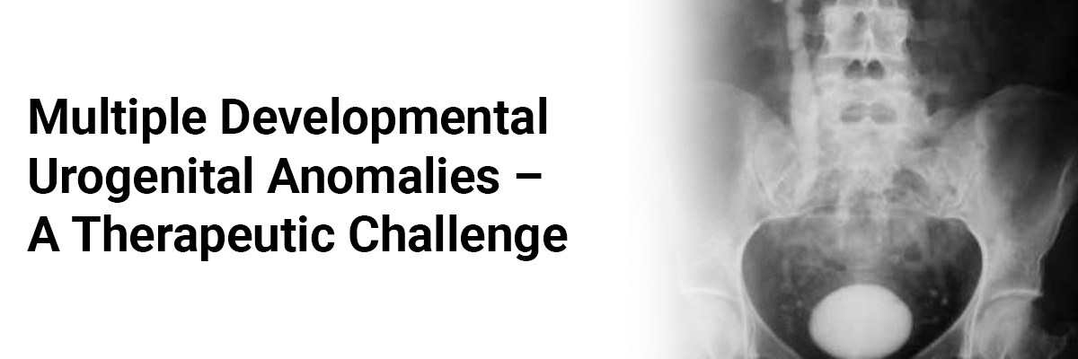
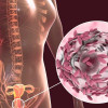
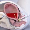
- A bicornuate uterus with hyperintense fluid accumulation in cervix and vagina confirming the hematocolpos.
Cystoscopy showed a hypospadiac meatus with a short urethra. The trigone appeared severely distorted and both ureteric orifices displaced laterally due to the pelvic mass. Further, a wide-open left ureteric orifice entered the diverticulum of the bladder. Further examination showed an atretic distal vagina and a huge globular mass palpable (rectally) above the atretic vaginal segment.
Four months later, the physicians performed a surgical exploration and considered a staged surgical protocol given multiple developmental urogenital anomalies. They made a "U" shaped incision between the perineum and hypospadiac meatus. They required a sharp dissection through the hard fibrotic tissue for a distance of 5cm from the fourchette to access the hematocolpos. Per rectal finger guidance facilitated the dissection.
The proximity of the bladder diverticulum with insertion of the left ureter into it indicated the possibility of accidental injury to the bladder or lower ureter during the intervention. A preoperative retrograde ureteric stenting was carried out to protect the left ureter, which failed due to extensive anatomical distortion by the mass.
Guidance with foley's catheter facilitated dissection without injuring the urinary tract. The hematocolpos was drained by incising its thick fibrotic wall and drained about 300ml of altered blood.
Postoperatively she was relieved of abdominal pain and resumed her first menstruation 45 days after the surgery. Re-examination during menstruation confirmed the patency of the vagina. A postoperative micturating cystogram showed no VUR on the left side.
Rajamaheswari N, Agarwal Sugandha, Chhikara Archana Bharti, Seethalakshmi K. Multiple Developmental Urogenital Anomalies: A Therapeutic Challenge. Indian Journal of Clinical Practice. 2012 Mar; 22(10): 529-531.
IJCP Editorial Team
Comprising seasoned professionals and experts from the medical field, the IJCP editorial team is dedicated to delivering timely and accurate content and thriving to provide attention-grabbing information for the readers. What sets them apart are their diverse expertise, spanning academia, research, and clinical practice, and their dedication to upholding the highest standards of quality and integrity. With a wealth of experience and a commitment to excellence, the IJCP editorial team strives to provide valuable perspectives, the latest trends, and in-depth analyses across various medical domains, all in a way that keeps you interested and engaged.
More FAQs by IJCP Editorial Team


Recent FAQs

Related FAQs


Medtalks is India's fastest growing Healthcare Learning and Patient Education Platform designed and developed to help doctors and other medical professionals to cater educational and training needs and to discover, discuss and learn the latest and best practices across 100+ medical specialties. Also find India Healthcare Latest Health News & Updates on the India Healthcare at Medtalks
Please login to comment on this article