 IJCP Editorial Team
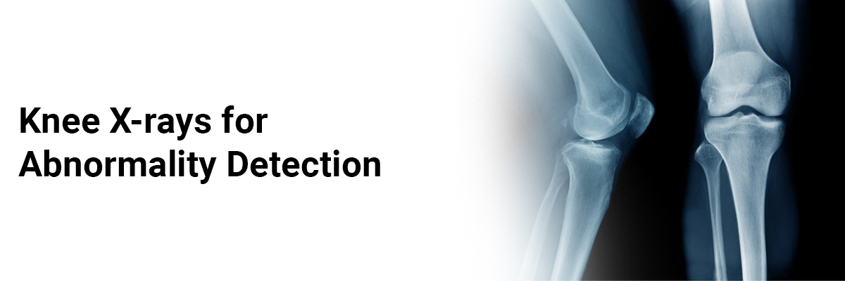
Knee X-rays for Abnormality Detection
Introduction
X-Ray is a commonly used diagnostic tool that produces an image of the internal anatomy of a person. It helps to identify and evaluate various medical conditions, from broken bones to more complex diseases. X-rays are a type of radiation that can pass through the body, creating images of the structures inside. This technology is essential for providing insight into a patient's health and detecting potential problems.
X-rays work by using a small amount of radiation to pass through the body and create a picture of the body's internal structures. The X-ray radiation passes through most of the body's soft tissues, such as skin, muscles, and organs, but is absorbed by denser materials, such as bones. The absorbed radiation creates a pattern of lighter and darker areas on an X-ray film or detector, which a radiologist interprets to diagnose any abnormalities.
Knee X-Ray
Healthcare providers may advise a knee X-ray to diagnose and treat any health issues concerning the knee or knees. A knee X-ray is a diagnostic procedure that generates a black-and-white image of the interior of the knee joint. This image will display the soft tissues and bones in and around the knee, such as the kneecap (patella), portion of the thigh bone (femur), and section of the shin bone (tibia). In some cases, a portion of the calf bone (fibula) also may be visible.
Need for Knee X-ray
An X-ray should be considered in any patient with knee pain or instability, as it can provide important diagnostic information and guide treatment options.
A knee X-ray is essential to detect underlying issues as it provides diagnostic information to identify any damage, disease, or other pathologies affecting the knee joint. Additionally, an X-ray can be used to assess the alignment of the joint and evaluate the integrity of the bone and soft tissue structures. Knee X-rays can be helpful in detecting:
• Broken knee bones or fractures.
• Joint dislocation.
• Excess fluid (a sign of a sprain).
• Loose fragments of bone.
• Bone spurs (or osteophytes).
• Osteoarthritis.
• Abnormal alignment of the knee joint.
• Bone infections (or osteomyelitis).
• Bone thinning (or osteopenia).
• Bone cancer.
Knee X-rays are also performed after a broken bone has been set to ensure that the bone or joint has healed adequately. In addition, in knee replacement cases, routine follow-up X-rays will help monitor the condition.
Sometimes healthcare providers may advise X-rays for both knees, which is called a bilateral X-ray and is usually done when the healthcare provider is looking for the signs of arthritis.
A knee X-ray may show-
• Soft-tissue changes: Apart from bones, X-ray images can also depict signs of soft-tissue swelling and excess fluid within the knee.
• Bone quality: X-rays independently can't evaluate bone density. However, they can detect abnormalities (like certain bone disorders and bone thinning).
• Alignment: X-ray images can depict the alignment of the knee joint and its abnormalities. Malalignment can exert excessive force on joint parts and accelerate arthritic changes.
• Joint spaces: The space between the knee bones is filled completely with cartilage. Narrowing this joint space is the best sign of the extent of knee arthritis.
• Early arthritis signs: X-ray images can also show other symptoms of arthritis, including bone spurs. Some of these early signs indicate the amount of knee pain due to early arthritis.
• Trauma/fracture: X-ray images also show evidence of injury to the bone, including fractures. Not all fractures may be visible on X-ray, but most do.
Sometimes your healthcare provider may advise additional images, and you may have to return for follow-up X-rays. They'll use these extra images to formulate an accurate diagnosis.
The procedure
A Knee X-ray is a simple and painless procedure carried out by a trained radiologic technologist, also known as an X-ray technician. Prior to the scan, patients should remove any jewelry or metal pieces from the vicinity of the area of interest. The scan itself typically takes around ten minutes and requires the patient to remain still for a few seconds until a beep is heard, at which point the X-ray technician will ask the patient to move. In certain circumstances, Magnetic Resonance Imaging (MRI) can be used in conjunction with a Knee X-ray to give a holistic view of any knee-related issues causing pain. It is essential that pregnant women must inform the doctor before undergoing an X-ray procedure.
Analysing an X-Ray - After the completion of the procedure, the report is sent to the medical practitioner for analysis. They assess the images to decide on the most suitable treatment plan. Afterward, the doctor evaluates the results compared to a standard knee to detect any irregularities.
Safety aspects - X-rays are generally considered safe, but there is the potential for developing cancer or harming the unborn baby in the womb. To ensure safety, doctors use the lowest possible radiation dose. It is advisable that pregnant women must protect themselves from the potential risk of X-rays by wearing a lead apron to minimize exposure.
The bottom line
An X-ray may be used to determine the condition of your knee, and an MRI may be necessary in some cases for further diagnosis. Although an MRI is a more advanced imaging test than an X-ray, an X-ray holds the advantage of being more useful in specific circumstances and a cost-effective technique. If you suspect any issues with your knee, it is best to consult a healthcare provider.
IJCP Editorial Team
Comprising seasoned professionals and experts from the medical field, the IJCP editorial team is dedicated to delivering timely and accurate content and thriving to provide attention-grabbing information for the readers. What sets them apart are their diverse expertise, spanning academia, research, and clinical practice, and their dedication to upholding the highest standards of quality and integrity. With a wealth of experience and a commitment to excellence, the IJCP editorial team strives to provide valuable perspectives, the latest trends, and in-depth analyses across various medical domains, all in a way that keeps you interested and engaged.
More FAQs by IJCP Editorial Team


.png)


Medtalks is India's fastest growing Healthcare Learning and Patient Education Platform designed and developed to help doctors and other medical professionals to cater educational and training needs and to discover, discuss and learn the latest and best practices across 100+ medical specialties. Also find India Healthcare Latest Health News & Updates on the India Healthcare at Medtalks
Please login to comment on this article