 IJCP Editorial Team
Embolization to Treat Uterine Fibroids with Bleeding and Severe Anemia
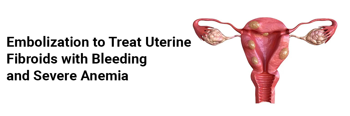
A report describes a case of a 35-year-old woman, para 3, who presented with acute menorrhagia. She described a history of three prior cesarean sections, with the latest one a year earlier. Before this episode, she reported regular menstrual cycles.
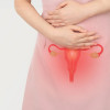
The examination was unremarkable, with a bulky uterus. Investigations revealed a negative urine pregnancy test, a hemoglobin level of 5.3 g/dL with normal platelet counts, and a normal pelvic ultrasound along with a follicle.
She reported a similar episode of menorrhagia two weeks before demanding admission to a local hospital, where she underwent curettage and received five units of packed cells.
Histology demonstrated benign endometrial cells. The menorrhagia episode recurred a week later, and she was readmitted, transfused four units of packed cells, and given an intrauterine balloon tamponade to arrest bleeding. She then got transferred to another hospital, where she received intramuscular progesterone 100 mg, maintenance oral progesterone, and intravenous antibiotics, along with packed cells and platelets. Her menorrhagia, however, persisted.
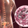
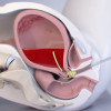
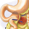
Investigations for hematological causes were negative. CT-pelvis revealed no arteriovenous malformation. She was initiated intravenous tranexamic acid and subsequently on oral estrogen when the bleeding persisted. Despite that, the bleeding continued, prompting pelvic angiogram and embolization of bilateral uterine arteries, which settled the bleeding, and then she received combined oral contraceptives (COC) before her discharge.
However, the patient presented again to the hospital a year later with prolonged heavy vaginal bleeding for 5 days. Before this, her menses were regular with normal menstrual flow. Adenomyosis was ruled-out. She received oral progesterone and got discharged the next day. Her hemoglobin on discharge was 12 g/dL.
She then re-presented the next day with severe menorrhagia complicated by symptomatic anemia. Her hemoglobin level saw a drop of 3 g/dL. She received blood transfusions, oral estrogen, and intravenous tranexamic acid. Despite these conservative measures, her vaginal bleeding persisted.
She underwent a pelvic angiogram and embolization of bilateral uterine arteries. Intraoperatively, she showed a tortuous right uterine artery with no arteriovenous malformation. Her bleeding settled, and she got discharged.
After discharge, the patient experienced an elective hysteroscopy with dilation and curettage, which revealed a normal endocervical and endometrial cavity. Histologic study of the endometrial curetting showed late secretory endometrium without hyperplasia or malignancy. The patient is now considering a hysterectomy.
Kho CL, Mathur M. Uterine artery embolisation for acute dysfunctional uterine bleeding with failed medical therapy: a novel approach to management. BMJ Case Rep. 2015 Jan 16;2015:bcr2014204446. doi: 10.1136/bcr-2014-204446. PMID: 25596287; PMCID: PMC4307078.
IJCP Editorial Team
Comprising seasoned professionals and experts from the medical field, the IJCP editorial team is dedicated to delivering timely and accurate content and thriving to provide attention-grabbing information for the readers. What sets them apart are their diverse expertise, spanning academia, research, and clinical practice, and their dedication to upholding the highest standards of quality and integrity. With a wealth of experience and a commitment to excellence, the IJCP editorial team strives to provide valuable perspectives, the latest trends, and in-depth analyses across various medical domains, all in a way that keeps you interested and engaged.
More FAQs by IJCP Editorial Team


Recent FAQs

Related FAQs


Medtalks is India's fastest growing Healthcare Learning and Patient Education Platform designed and developed to help doctors and other medical professionals to cater educational and training needs and to discover, discuss and learn the latest and best practices across 100+ medical specialties. Also find India Healthcare Latest Health News & Updates on the India Healthcare at Medtalks
Please login to comment on this article