 IJCP Editorial Team
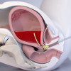
Diagnostic Accuracy of Female Pelvic Ultrasonography in Differentiating Precocious Puberty from Premature Thelarche: A Systematic Review and Meta-analysis
Discrimination between precocious puberty (PP) and premature thelarche (PT) is difficult. Hormonal testing (i.e., the GnRH stimulation test) is considered the gold standard for diagnosing precocious puberty (PP). However, it is invasive, time-consuming, costly, and may create an unpleasant experience for participants. The relatively low sensitivity of this test, despite its high specificity, is another significant drawback. This is primarily due to the insufficient luteinizing hormone (LH)-response to the GnRH in the early stages of premature sexual development. This hormonal test's diagnostic utility is thus constrained. A group of researchers conducted a systematic review and meta-analysis to compare the pelvic ultrasonography parameters between PP and PT patients and determine its usefulness in the diagnostic accuracy of these parameters compared with the GnRH stimulation test. Data was sourced from PubMed/MEDLINE, EMBASE, Scopus, and Cochrane Library databases from inception up to March 31, 2021. All types of studies were included except for case reports and review articles. Those whose organic conditions might cause PP were excluded. Out of 3,273 articles from the above-mentioned databases, 13 studies were included in this systematic review and meta-analysis. The mean, standard deviation, sensitivity, and specificity of each parameter were documented. Forest plots were constructed to display the estimated standardized mean differences (SMDs) from each included study and the overall calculations. A bivariate model was used to calculate the pooled sensitivity, specificity, positive likelihood ratio (PLR), negative likelihood ratio (NLR), and diagnostic odds ratio (DOR).
The SMDs (95% confidence interval – CI) in ovarian volume, fundal-cervical ratio (FCR), uterine length, uterine cross-sectional area(CSA), and uterine volume between PP and PT groups were 1.12 (0.78–1.45; p < 0.01), 0.90 (0.07–1.73; p = 0.03), 1.38 (0.99–1.78; p < 0.01), 1.06 (0.61–1.50; p < 0.01), and 1.21 (0.84–1.58; p <0.01), respectively. A uterine length of 3.20 cm yielded a pooled sensitivity of 81.8% (95% CI 78.3%–84.9%), specificity of 82.0% (95% CI 61.0%–93.0%), PLR of 4.56 (95% CI 2.15–9.69), NLR of 0.26 (95% CI 0.17–0.39), and DOR of 19.62 (95% CI 6.45–59.68). All investigated ultrasonography parameters were significantly greater in the PP group than in the PT group. The early increases in uterine and ovarian sizes represent the estrogenic effects of hypothalamic-pituitary-gonadal (HPG) axis activation on internal female genitalia, which indicates PP. Hence, girls with PP had significantly greater uterine and ovarian measurements as compared to PT. Furthermore, uterine length represented a reliable marker to differentiate PP from PT, thereby reducing the possibility of misdiagnosing PP. Therefore, to improve diagnostic precision, pelvic ultrasonography emphasizing these measurements should be considered as an adjunct to clinical examination, bone radiography, and laboratory tests.
Nguyen, N N, et al. Front Endocrinol
(Lausanne). 2021 Septemer 1;12:735875.
IJCP Editorial Team
Comprising seasoned professionals and experts from the medical field, the IJCP editorial team is dedicated to delivering timely and accurate content and thriving to provide attention-grabbing information for the readers. What sets them apart are their diverse expertise, spanning academia, research, and clinical practice, and their dedication to upholding the highest standards of quality and integrity. With a wealth of experience and a commitment to excellence, the IJCP editorial team strives to provide valuable perspectives, the latest trends, and in-depth analyses across various medical domains, all in a way that keeps you interested and engaged.
More FAQs by IJCP Editorial Team


Recent FAQs

Related FAQs


Medtalks is India's fastest growing Healthcare Learning and Patient Education Platform designed and developed to help doctors and other medical professionals to cater educational and training needs and to discover, discuss and learn the latest and best practices across 100+ medical specialties. Also find India Healthcare Latest Health News & Updates on the India Healthcare at Medtalks
Please login to comment on this article