 IJCP Editorial Team
Application of Magnetic Resonance Imaging and Ultrasound Fusion Technology in Gynecology
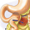
With the development of imaging methods, a fusion of MRI and Ultrasound data has grown in popularity, in particular in intestinal and prostate imaging. This technique of fusion allows optimization of biopsy samples by facilitating a targeted approach broadening the diagnostic possibilities in non-invasive ways. The use and storage of digital data allow immediate review ofinformation obtained. However, the MRI-Ultrasound fusion method is underdeveloped in the gynecology field. Combining these two methods may improve the detection of abnormalities above the range of individual method applications.
The MRI-Ultrasound technique can be performed on the same day to ensure accurate synchronization of the pelvic floor. The method can be performed by a radiologist or by a gynecologist assisted by a radiologist with an MRI examination performed first. The MRI data can be then transferred from the Picture Archiving Communication System (PACS), or, failing that, from a CD ROM (recordable compact disc) to the ultrasound machine and the Ultrasound images can be obtained from LOGIQ E10(GE Healthcare) machine. However, there are certain pre-requisites before an MRI scan such as fasting 3 hours before the scan, emptying the bladder before taking the images, etc. AfterMRI, care must be taken to ensure the patient doesn’t develop hypoglycemia that may interfere with ultrasound-MRI fusion.
The success of the fusion depends on the precision of the images, so MRI and ultrasound have the same spatial coordinates for each modality. This can be achieved when imaging a small stationary object such as the prostate, however, it would be challenging for the pelvic floor in automatic as well as manual mode. The lack of recognized landmarks for synchronization on the patient’s abdominal wall has prevented the implementation of the method on a large scale and represents a major limitation in the gynecology field.
A preliminary study has shown that the fusion method can detect adenomyosis, but the lack of standards limits its applicability. Several other studies have suggested a potential function of fusion imaging in identifying gynecological cancers. These studies suggested that fusion imaging is feasible in patients with local cervical and ovarian cancer and may increase the accuracy relative to the individual imaging methods. However, further studies are required to standardize the new tool for gynecological diseases and to assess its functionality.
Source:
Bazot M et al., Ultrasound in Obstetrics &
Gynecology, Volume 59, Issue 2, p. 141-14,https://doi.org/10.1002/uog.24754
IJCP Editorial Team
Comprising seasoned professionals and experts from the medical field, the IJCP editorial team is dedicated to delivering timely and accurate content and thriving to provide attention-grabbing information for the readers. What sets them apart are their diverse expertise, spanning academia, research, and clinical practice, and their dedication to upholding the highest standards of quality and integrity. With a wealth of experience and a commitment to excellence, the IJCP editorial team strives to provide valuable perspectives, the latest trends, and in-depth analyses across various medical domains, all in a way that keeps you interested and engaged.
More FAQs by IJCP Editorial Team


Recent FAQs

Related FAQs


Medtalks is India's fastest growing Healthcare Learning and Patient Education Platform designed and developed to help doctors and other medical professionals to cater educational and training needs and to discover, discuss and learn the latest and best practices across 100+ medical specialties. Also find India Healthcare Latest Health News & Updates on the India Healthcare at Medtalks
Please login to comment on this article