Acute Fatty Liver of Pregnancy in a 28-Year-Old Primigravida at 35 Weeks Gestation
A report describes a case of a 28-year-old primigravida at 35 weeks of gestation who presented with severe nausea, vomiting, and significant fatigue for five days. Her antenatal care had been up-to-date, and her first two trimesters were largely uneventful apart from mild anemia and vomiting during the early first trimester. An ultrasound two weeks prior showed normal physiological changes, with normal liver and kidney scans. Mild hydronephrosis and dilated ureters were noted, consistent with normal pregnancy changes. However, amniotic fluid assessment revealed a low amniotic fluid index (AFI) of 3 cm, below the typical range of 5–25 cm, indicating oligohydramnios.
On examination, the patient exhibited pedal edema, icterus, and elevated blood pressure (138/80 mmHg). She had no significant medical history and tested negative for hepatitis B, C, E, and human immunodeficiency virus (HIV). She was also not a previous chronic aspirin or paracetamol user.
Ultrasound at this visit showed an AFI of 1–2 cm, indicating severe oligohydramnios, along with a slightly enlarged liver exhibiting fatty changes. Laboratory investigations revealed dimorphic red blood cells with marked anisopoikilocytosis, leukocytosis, abnormal liver and renal function tests, and an altered lipid profile. The patient was euglycemic but hypertensive (140/80 mmHg) and had a low Bishop’s score of 3. Given these findings, she consented to an emergency lower-segment cesarean section (LSCS).
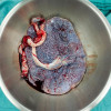
Before surgery, she received transfusions including two units of cryoprecipitate, four units of frozen plasma, and four platelet transfusions. LSCS was performed without complications, and post-delivery laboratory tests confirmed anemia, coagulopathy, and persistent liver and renal function abnormalities, as shown in Table 1 below.
|
Lab investigations |
Patient’s values |
Normal Range |
|
Hemoglobin (g/dL) |
8.4 |
9.5 – 15 |
|
Total Leucocyte count (×109/L) |
20.09 |
6 – 16 |
|
Platelet (×109/L) |
179 |
145 – 400 |
|
Blood Urea (mg/dL) |
10.7 |
5 – 20 |
|
Uric Acid (mg/dL) |
6.4 |
2.4 – 6.0 |
|
Creatinine (mg/dL) |
1.19 |
0.4 – 0.8 |
|
Serum LDH (U/L) |
774 |
82 – 524
|
|
Plasma Na+ (mmol/L) |
134 |
130 – 140 |
|
Plasma K+ (mmol/L) |
4.52 |
3.3 – 5.1 |
|
Blood Ammonia (µg/dL) |
130.8 |
19 – 60 |
|
ALT (U/L) |
168 |
10 – 49 |
|
AST (U/L) |
214 |
0 – 34 |
|
Serum Alkaline Phosphatase (U/L) |
264 |
0 – 129 |
|
Gamma Glutamyl Transferase (U/L) |
664 |
5 – 55 |
|
Total Bilirubin (mg/dL) |
8.70 |
0.3 – 1.2 |
|
Direct Bilirubin (mg/dL) |
7.16 |
0 – 0.3 |
|
Indirect Bilirubin (mg/dL) |
1.54 |
0.1 – 0.5 |
|
Serum Protein (g/dL) |
4.54 |
5.7 – 8.2 |
|
Serum Albumin (g/dL) |
2.29 |
3.2 – 4.8 |
|
Albumin/Globulin Ratio |
1.02 |
1.5 – 2.5 |
|
PT (seconds) |
42.9 |
8.6 – 12.4 |
|
APTT (seconds) |
69.7 |
26 – 35 |
|
D-DIMER (µg/mL)3 |
20.1 |
0.14 – 2.82 |
|
Serum Protein (g/dL) |
4.54 |
5.7 – 8.2
|
|
Serum Albumin (g/dL) |
2.29 |
3.2 – 4.8 |
|
Serum Globulin (g/dL) |
2.25 |
2 – 3.5 |
PT: Prothrombin time, APTT: Activated partial thromboplastin time, ALT: Alanine aminotransferase, AST: Aspartate aminotransferase
Table 1: Laboratory Test Post Delivery
Both mother and child
were monitored in the intensive care unit for a week. Follow-up lipid profile
revealed severely low High-Density Lipoprotein (HDL) levels, elevated Very
Low-Density Lipoprotein (VLDL), and an increased cholesterol-to-HDL ratio as
shown in Table 2 below.
|
Lab investigations |
Patient’s values |
Normal Range |
|
Cholesterol (mg/dL) |
106.0 |
110 – 200 |
|
HDL (mg/dL) |
8.0 |
40 – 60 |
|
Triglycerides (mg/dL) |
282.0 |
40 – 200 |
|
VLDL (mg/dL) |
56.4 |
10 – 40 |
|
LDL (mg/dL) |
73.00 |
0 – 100 |
|
Cholesterol/HDL ratio |
13.25 |
0 – 4.1 |
Table 2: Follow-Up Lipid Profile
This case shows the importance of early recognition and prompt management of acute fatty liver of pregnancy (AFLP), a life-threatening condition. Rapid diagnosis, immediate hospital admission, and urgent delivery are imperative to prevent maternal and fetal morbidity and mortality.


Recent FAQs


Related FAQs


Medtalks is India's fastest growing Healthcare Learning and Patient Education Platform designed and developed to help doctors and other medical professionals to cater educational and training needs and to discover, discuss and learn the latest and best practices across 100+ medical specialties. Also find India Healthcare Latest Health News & Updates on the India Healthcare at Medtalks
Please login to comment on this article