A Case Report of Using MRI for Diagnosing Placenta Accreta
Placenta accreta spectrum (PAS) involves the abnormal attachment of the placental tissue to the uterine muscle. It can develop under various conditions and is primarily diagnosed using ultrasound. When ultrasound results are inconclusive, magnetic resonance imaging (MRI) is highly effective and aids in surgical planning. The definitive diagnosis is confirmed during surgery and by pathology.
A 35-year-old woman with a history of two cesarean sections and chronic hypertension presented with a 23-week pregnancy and obstetric ultrasound that revealed a totally occlusive anterior placenta, with some placental lacunae, thinning of the perivesical myometrium, and loss of a clear zone, consistent with placenta accreta.
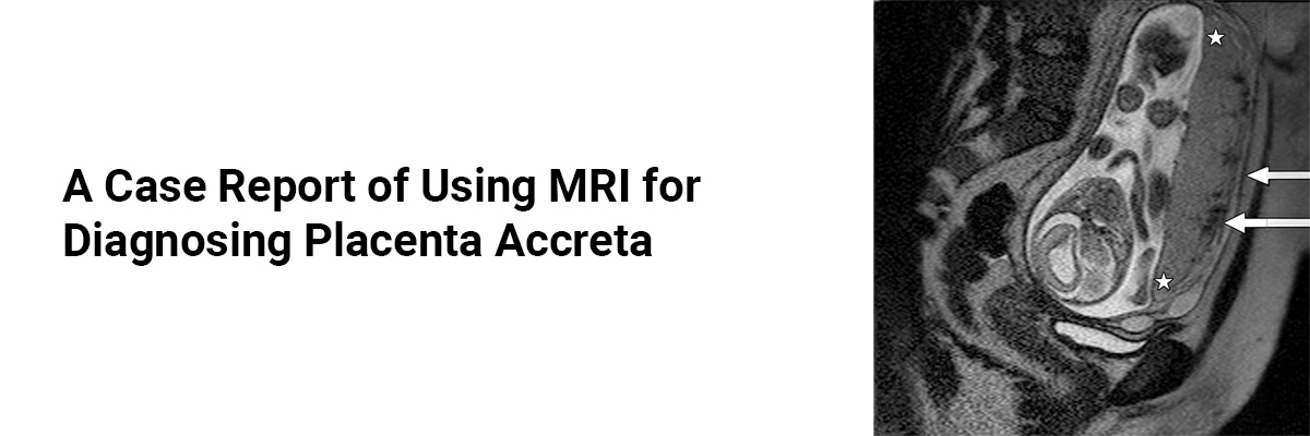
MRI conducted at 27 weeks of pregnancy, revealed showed uterine bulge, with an hourglass shape, anterior and low insertion placenta, determining occlusion of the internal cervical os, as well as low-signal bands on T2WI and some aberrant vascular structures. This confirmed the diagnosis of accreta, showing no extension to nearby organs. At 34 weeks, she underwent a cesarean section and hysterectomy, which revealed no bladder invasion.
To summarize, PAS is typically diagnosed antenatally via ultrasound, but MRI can provide additional, crucial details. Radiologists should be familiar with the characteristic MRI features of PAS.
Recently, the Society for Abdominal Radiology (SAR) and the European Society for Urogenital Radiology (ESUR) have jointly described seven MRI features of PAS disorders, that include the following;
Intraplacental T2-dark bands, which is the more sensitive feature for the diagnosis of PAS disorders • Placental or uterine bulge • Myometrial thinning • Bladder wall interruption • Focal exophytic mass • Loss of T2-hypointense retroplacental line, and • Abnormal vascularization of the placental bed.• Intraplacental T2-dark bands, which is the more sensitive feature for the diagnosis of PAS disorders
- Placental or uterine bulge
- Myometrial thinning
- Bladder wall interruption
- Focal exophytic mass
- Loss of T2-hypointense retroplacental line, and
- Abnormal vascularization of the placental bed.
Source: Aliaga F, del Campo F, Cocio R, Schiappacasse G, Blanco S, Pires Y. MRI and placenta accreta: Keys for its interpretation in images, regarding a case. J Case Rep Images Obstet Gynecol 2024;10(2):50–53.


Please login to comment on this article