 IJCP Editorial Team
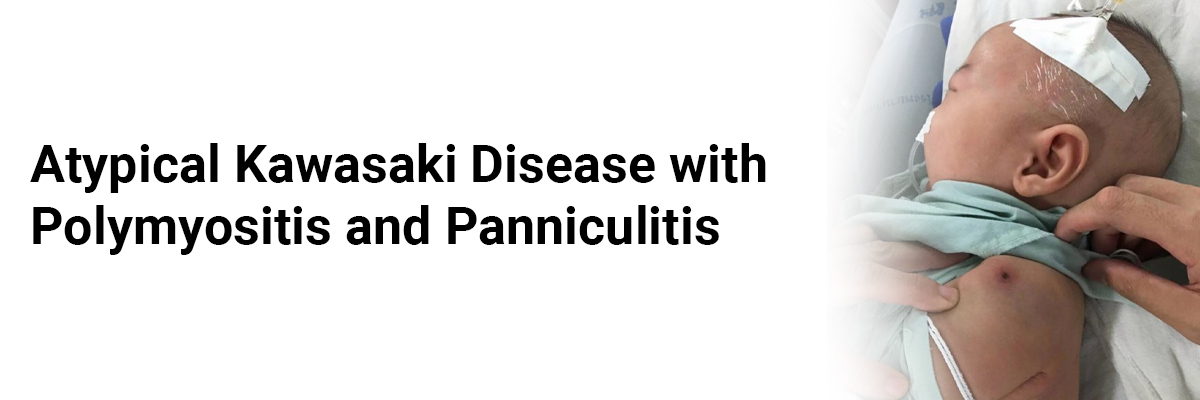
Atypical Kawasaki disease with polymyositis and panniculitis
A 3-year-old boy, complained of pain in both lower limbs for eight days and fever for seven days. He was apparently healthy and went to bed symptom-free and awoke in the morning with diffuse pain restricted to only both lower limbs. The pain severity resulted in him refusing to walk, with no loss of movement. The next day, he developed a moderate-degree fever, not associated with chills or rigors. There wasn't any history of local swelling in either lower limb, warmth, redness, or stiffness of his ankles, knees, or hips; or any rash, cough, coryza, sore throat, breathlessness, or palpitations.
Additionally, there wasn't any history of preceding trauma or bleeding from any site, progressive pallor, weight loss, or appetite. The child had regular Bowel and bladder habits, although he had difficulty squatting. His sensorium was normal but was irritable due to the pain.
His all other history was non-significant.
On admission, he was febrile (38.5°C), with tachycardia (pulse rate of 136/min), normal respiratory rate, blood pressure, and capillary refill time. His general physical examination was normal.
Musculoskeletal examination showed diffuse tenderness over bilateral extensor aspects of the thighs and legs, without erythema, swelling, or increased temperature of the joints. The physicians couldn't assess Localized joint tenderness or restricted range of motion of any joint due to the child's uncooperative behavior. However, a local examination of the spine was normal.
The motor component of the lower limb's central nervous system examination (CNS) showed normal bulk; however, the physicians did not assess the gait, power, tone, and deep tendon reflexes due to the considerable pain and tenderness. The remaining CNS, cardiovascular, respiratory, and abdominal examinations stood non-contributory.
The clinical phenotype of the child indicated the possibility of an infectious, inflammatory, or autoimmune etiopathogenesis, including acute polymyositis, acute osteomyelitis, septicemia, septic arthritis/reactive arthritis, and acute rheumatic fever (ARF).
Lab findings demonstrated an elevated erythrocyte sedimentation rate (ESR) of 105 mm at 1-h, neutrophilic leukocytosis with total leukocyte count (TLC) 25 × 109/L, a differential count of 75% neutrophils (N), 15% lymphocytes (L), 6% eosinophils (E), and 4% monocytes (M), mild anemia (hemoglobin 10.6 g/dL), normal platelet count (384 × 109/L), and a peripheral smear displaying normocytic normochromic red blood cells with absence of atypical or malignant cells. These findings suggested the acute infectious differential diagnoses of acute polymyositis or acute osteomyelitis. Thus, the patient was started intravenous cloxacillin and oral ibuprofen, pending further investigation reports. The X-rays and ultrasonography reports were normal. Magnetic resonance imaging (MRI) of the lower limbs revealed diffuse patchy areas of hyperintensities in all the thigh muscles, involving all the compartments. However, serum creatine kinase (CK) and lactate dehydrogenase (LDH) levels were normal. A Technetium-99 bone scan, Liver and kidney function tests also proved to be normal. Two blood cultures conducted within a 24-h span were sterile.
After ruling out the acute infectious etiologies, and observing the persisting symptoms, the physicians considered other relevant diagnoses. They ruled out hepatolenticular malignancies and other infiltrative disorders; systemic-onset juvenile idiopathic arthritis (SOJIA), and acute pain crisis associated with sickle cell anemia by relevant tests.
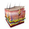
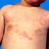
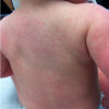
By the end of the 2nd week, the child developed periungual desquamation in the toes, without redness of lips, strawberry tongue, conjunctival congestion, edema of the dorsal aspect of hands and feet, rash, and significant lymphadenopathy. He also developed painless, subcutaneous nodules on the left forearm's extensor aspect and the right leg's shin, which was confirmed as panniculitis by biopsy. Repeat lab findings showed increasing leukocytosis (TLC 35.25 × 10⁹/L with 73% N, 18% L, 5% E, and 4% M), thrombocytosis (690 × 109/L), and a further rise in ESR (120 mm at 1-h). These indicated the possibility of atypical KD and multisystem inflammatory syndrome in children. Reverse transcription polymerase chain reaction and antibodies for coronavirus disease- 2019 were negative. An echocardiogram showed a thin rim of pericardial effusion and dilatation of the left main coronary artery (LCA) with 2.55 Z (normal cut-off <2 Z score), thereby satisfying the diagnostic criteria for atypical Kawasaki disease (KD).
Hence, the clinicians diagnosed him with atypical KD on the 13th day of illness. They initiated intravenous immunoglobulin (IVIg) in a single dose of 2 g/kg with aspirin (75 mg/kg/day) in the child. No defervescence within 72 h of the first dose prompted a trial of the second dose of IVIg. After this treatment, the child recovered from fever and limb pain, and by the 5th day, he had become ambulatory.
The physicians discharged him on low-dose aspirin (5 mg/kg/day).
On follow-up, four weeks later, he remained symptomless.
However, they prescribed clopidogrel (1 mg/kg/day) along with the low-dose aspirin as his repeat echocardiography showed persistent dilatation of the LCA.
Rajasegar R, Sugumar K, Chandrasekaran V, Gunasekaran D, Anantharaj A. Atypical Kawasaki disease with polymyositis and panniculitis: Case report and review of literature. Indian Pediatr Case Rep 2022;2:79-83
IJCP Editorial Team
Comprising seasoned professionals and experts from the medical field, the IJCP editorial team is dedicated to delivering timely and accurate content and thriving to provide attention-grabbing information for the readers. What sets them apart are their diverse expertise, spanning academia, research, and clinical practice, and their dedication to upholding the highest standards of quality and integrity. With a wealth of experience and a commitment to excellence, the IJCP editorial team strives to provide valuable perspectives, the latest trends, and in-depth analyses across various medical domains, all in a way that keeps you interested and engaged.
More FAQs by IJCP Editorial Team


Recent FAQs


Related FAQs


Medtalks is India's fastest growing Healthcare Learning and Patient Education Platform designed and developed to help doctors and other medical professionals to cater educational and training needs and to discover, discuss and learn the latest and best practices across 100+ medical specialties. Also find India Healthcare Latest Health News & Updates on the India Healthcare at Medtalks
Please login to comment on this article